The sequence of sequencers: The history of sequencing DNA_the coding sequence of-程序员宅基地
The sequence of sequencers: The history of sequencing DNA
Author links open overlay panelJames M.HeatherBenjaminChain
Show moreAdd to Mendeley
Share
Cite
https://doi.org/10.1016/j.ygeno.2015.11.003Get rights and content
Under an Elsevier user license
open archive
Highlights
•
We review the drastic changes to DNA sequencing technology over the last 50 years.
•
First-generation methods enabled sequencing of clonal DNA populations.
•
The second-generation massively increased throughput by parallelizing many reactions.
•
Third-generation methods allow direct sequencing of single DNA molecules.
Abstract
Determining the order of nucleic acid residues in biological samples is an integral component of a wide variety of research applications. Over the last fifty years large numbers of researchers have applied themselves to the production of techniques and technologies to facilitate this feat, sequencing DNA and RNA molecules. This time-scale has witnessed tremendous changes, moving from sequencing short oligonucleotides to millions of bases, from struggling towards the deduction of the coding sequence of a single gene to rapid and widely available whole genome sequencing. This article traverses those years, iterating through the different generations of sequencing technology, highlighting some of the key discoveries, researchers, and sequences along the way.
Keywords
DNA
RNA
Sequencing
Sequencer
History
1. Introduction
“... [A] knowledge of sequences could contribute much to our understanding of living matter.”
Frederick Sanger [1]
The order of nucleic acids in polynucleotide chains ultimately contains the information for the hereditary and biochemical properties of terrestrial life. Therefore the ability to measure or infer such sequences is imperative to biological research. This review deals with how researchers throughout the years have addressed the problem of how to sequence DNA, and the characteristics that define each generation of methodologies for doing so.
2. First-generation DNA sequencing
Watson and Crick famously solved the three-dimensional structure of DNA in 1953, working from crystallographic data produced by Rosalind Franklin and Maurice Wilkins [2], [3], which contributed to a conceptual framework for both DNA replication and encoding proteins in nucleic acids. However, the ability to ‘read’ or sequence DNA did not follow for some time. Strategies developed to infer the sequence of protein chains did not seem to readily apply to nucleic acid investigations: DNA molecules were much longer and made of fewer units that were more similar to one another, making it harder to distinguish between them [4]. New tactics needed to be developed.
Initial efforts focused on sequencing the most readily available populations of relatively pure RNA species, such as microbial ribosomal or transfer RNA, or the genomes of single-stranded RNA bacteriophages. Not only could these be readily bulk-produced in culture, but they are also not complicated by a complementary strand, and are often considerably shorter than eukaryotic DNA molecules. Furthermore, RNase enzymes able to cut RNA chains at specific sites were already known and available. Despite these advantages, progress remained slow, as the techniques available to researchers – borrowed from analytical chemistry – were only able to measure nucleotide composition, and not order [5]. However, by combining these techniques with selective ribonuclease treatments to produce fully and partially degraded RNA fragments [6] (and incorporating the observation that RNA contained a different nucleotide base [7]), in 1965 Robert Holley and colleagues were able to produce the first whole nucleic acid sequence, that of alanine tRNA from Saccharomyces cerevisiae [8]. In parallel, Fred Sanger and colleagues developed a related technique based on the detection of radiolabelled partial-digestion fragments after two-dimensional fractionation [9], which allowed researchers to steadily add to the growing pool of ribosomal and transfer RNA sequences [10], [11], [12], [13], [14]. It was also by using this 2-D fractionation method that Walter Fiers' laboratory was able to produce the first complete protein-coding gene sequence in 1972, that of the coat protein of bacteriophage MS2 [15], followed four years later by its complete genome [16].
It was around this time that various researchers began to adapt their methods in order to sequence DNA, aided by the recent purification of bacteriophages with DNA genomes, providing an ideal source for testing new protocols. Making use of the observation that Enterobacteria phage λ possessed 5′ overhanging ‘cohesive’ ends, Ray Wu and Dale Kaiser used DNA polymerase to fill the ends in with radioactive nucleotides, supplying each nucleotide one at a time and measuring incorporation to deduce sequence [17], [18]. It was not long before this principle was generalized through the use of specific oligonucleotides to prime the DNA polymerase. Incorporation of radioactive nucleotides could then be used to infer the order of nucleotides anywhere, not just at the end termini of bacteriophage genomes [19], [20], [21]. However the actual determination of bases was still restricted to short stretches of DNA, and still typically involved a considerable amount of analytical chemistry and fractionation procedures.
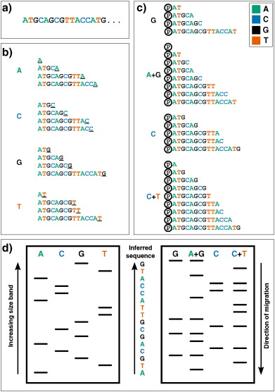
The next practical change to make a large impact was the replacement of 2-D fractionation (which often consisted of both electrophoresis and chromatography) with a single separation by polynucleotide length via electrophoresis through polyacrylamide gels, which provided much greater resolving power. This technique was used in two influential yet complex protocols from the mid-1970s: Alan Coulson and Sanger's ‘plus and minus’ system in 1975 and Allan Maxam and Walter Gilbert's chemical cleavage technique [22], [23]. The plus and minus technique used DNA polymerase to synthesize from a primer, incorporating radiolabelled nucleotides, before performing two second polymerisation reactions: a ‘plus’ reaction, in which only a single type of nucleotide is present, thus all extensions will end with that base, and a ‘minus’ reaction, in which three are used, which produces sequences up to the position before the next missing nucleotide. By running the products on a polyacrylamide gel and comparing between the eight lanes, one is able to infer the position of nucleotides at each position in the covered sequence (except for those which lie within a homopolymer, i.e. a run of the same nucleotide). It was using this technique that Sanger and colleagues sequenced the first DNA genome, that of bacteriophage ϕX174 (or ‘PhiX’, which enjoys a position in many sequencing labs today as a positive control genome) [24]. While still using polyacrylamide gels to resolve DNA fragments, the Maxam and Gilbert technique differed significantly in its approach. Instead of relying on DNA polymerase to generate fragments, radiolabelled DNA is treated with chemicals which break the chain at specific bases; after running on a polyacrylamide gel the length of cleaved fragments (and thus position of specific nucleotides) can be determined and therefore sequence inferred (see Fig. 1, right). This was the first technique to be widely adopted, and thus might be considered the real birth of ‘first-generation’ DNA sequencing.
Fig. 1. First-generation DNA sequencing technologies. Example DNA to be sequenced (a) is illustrated undergoing either Sanger (b) or Maxam–Gilbert (c) sequencing. (b): Sanger's ‘chain-termination’ sequencing. Radio- or fluorescently-labelled ddNTP nucleotides of a given type - which once incorporated, prevent further extension - are included in DNA polymerisation reactions at low concentrations (primed off a 5′ sequence, not shown). Therefore in each of the four reactions, sequence fragments are generated with 3′ truncations as a ddNTP is randomly incorporated at a particular instance of that base (underlined 3′ terminal characters). (c): Maxam and Gilbert's ‘chemical sequencing’ method. DNA must first be labelled, typically by inclusion of radioactive P 32 in its 5′ phosphate moiety (shown here by Ⓟ). Different chemical treatments are then used to selectively remove the base from a small proportion of DNA sites. Hydrazine removes bases from pyrimidines (cytosine and thymine), while hydrazine in the presence of high salt concentrations can only remove those from cytosine. Acid can then be used to remove the bases from purines (adenine and guanine), with dimethyl sulfate being used to attack guanines (although adenine will also be affected to a much lesser extent). Piperidine is then used to cleave the phophodiester backbone at the abasic site, yielding fragments of variable length. (d): Fragments generated from either methodology can then be visualized via electrophoresis on a high-resolution polyacrylamide gel: sequences are then inferred by reading ‘up’ the gel, as the shorter DNA fragments migrate fastest. In Sanger sequencing (left) the sequence is inferred by finding the lane in which the band is present for a given site, as the 3′ terminating labelled ddNTP corresponds to the base at that position. Maxam–Gilbert sequencing requires a small additional logical step: Ts and As can be directly inferred from a band in the pyrimidine or purine lanes respectively, while G and C are indicated by the presence of dual bands in the G and A + G lanes, or C and C + T lanes respectively.
However the major breakthrough that forever altered the progress of DNA sequencing technology came in 1977, with the development of Sanger's ‘chain-termination’ or dideoxy technique [25]. The chain-termination technique makes use of chemical analogues of the deoxyribonucleotides (dNTPs) that are the monomers of DNA strands. Dideoxynucleotides (ddNTPs) lack the 3′ hydroxyl group that is required for extension of DNA chains, and therefore cannot form a bond with the 5′ phosphate of the next dNTP [26]. Mixing radiolabelled ddNTPs into a DNA extension reaction at a fraction of the concentration of standard dNTPs results in DNA strands of each possible length being produced, as the dideoxy nucleotides get randomly incorporated as the strand extends, halting further progression. By performing four parallel reactions containing each individual ddNTP base and running the results on four lanes of a polyacrylamide gel, one is able to use autoradiography to infer what the nucleotide sequence in the original template was, as there will a radioactive band in the corresponding lane at that position of the gel (see Fig. 1, left). While working on the same principle as other techniques (that of producing all possible incremental length sequences and labelling the ultimate nucleotide), the accuracy, robustness and ease of use led to the dideoxy chain-termination method – or simply, Sanger sequencing – to become the most common technology used to sequence DNA for years to come.
A number of improvements were made to Sanger sequencing in the following years, which primarily involved the replacement of phospho- or tritrium-radiolabelling with fluorometric based detection (allowing the reaction to occur in one vessel instead of four) and improved detection through capillary based electrophoresis. Both of these improvements contributed to the development of increasingly automated DNA sequencing machines [27], [28], [29], [30], [31], [32], [33], and subsequently the first crop of commercial DNA sequencing machines [34] which were used to sequence the genomes of increasingly complex species.
These first-generation DNA sequencing machines produce reads slightly less than one kilobase (kb) in length: in order to analyse longer fragments researchers made use of techniques such as ‘shotgun sequencing’ where overlapping DNA fragments were cloned and sequenced separately, and then assembled into one long contiguous sequence (or ‘contig’) in silico [35], [36]. The development of techniques such as polymerase chain reaction (PCR) [37], [38] and recombinant DNA technologies [39], [40] further aided the genomics revolution by providing means of generating the high concentrations of pure DNA species required for sequencing. Improvements in sequencing also occurred by less direct routes. For instance, the Klenow fragment DNA polymerase – a fragment of the Escherichia coli DNA polymerase that lacks 5′ to 3′ exonuclease activity, produced through protease digestion of the native enzyme [41] – had originally been used for sequencing due to its ability to incorporate ddNTPs efficiently. However, more sequenced genomes and tools for genetic manipulation provided the resources to find polymerases that were better at accommodating the additional chemical moeities of the increasingly modified dNTPs used for sequencing [42]. Eventually, newer dideoxy sequencers – such as the ABI PRISM range developed from Leroy Hood's research, produced by Applied Biosystems [43], which allowed simultaneous sequencing of hundreds of samples [44] – came to be used in the Human Genome Project, helping to produce the first draft of that mammoth undertaking years ahead of schedule [45], [46].
3. Second-generation DNA sequencing
Concurrent with the development of large-scale dideoxy sequencing efforts, another technique appeared that set the stage for the first wave in the next generation of DNA sequencers. This method markedly differed from existing methods in that it did not infer nucleotide identity through using radio- or fluorescently-labelled dNTPs or oligonucleotides before visualising with electrophoresis. Instead researchers utilized a recently discovered luminescent method for measuring pyrophosphate synthesis: this consisted of a two-enzyme process in which ATP sulfurylase is used to convert pyrophosphate into ATP, which is then used as the substrate for luciferase, thus producing light proportional to the amount of pyrophosphate [47]. This approach was used to infer sequence by measuring pyrophosphate production as each nucleotide is washed through the system in turn over the template DNA affixed to a solid phase [48]. Note that despite the differences, both Sanger's dideoxy and this pyrosequencing method are ‘sequence-by-synthesis’ (SBS) techniques, as they both require the direct action of DNA polymerase to produce the observable output (in contrast to the Maxam–Gilbert technique). This pyrosequencing technique, pioneered by Pål Nyrén and colleagues, possessed a number of features that were considered beneficial: it could be performed using natural nucleotides (instead of the heavily-modified dNTPs used in the chain-termination protocols), and observed in real time (instead of requiring lengthy electrophoreses) [49], [50], [51]. Later improvements included attaching the DNA to paramagnetic beads, and enzymatically degrading unincorporated dNTPs to remove the need for lengthy washing steps. The major difficulty posed by this technique is finding out how many of the same nucleotide there are in a row at a given position: the intensity of light released corresponds to the length of the homopolymer, but noise produced a non-linear readout above four or five identical nucleotides [51]. Pyrosequencing was later licensed to 454 Life Sciences, a biotechnology company founded by Jonathan Rothburg, where it evolved into the first major successful commercial ‘next-generation sequencing’ (NGS) technology.
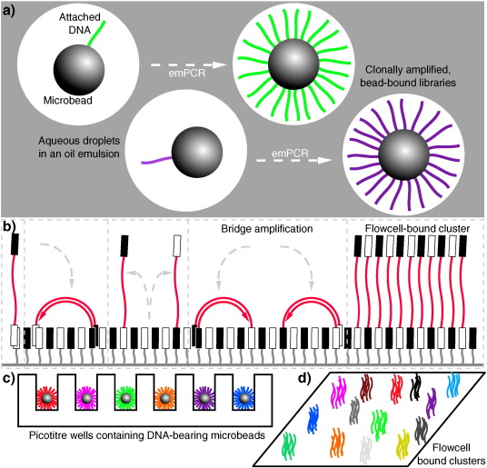
The sequencing machines produced by 454 (later purchased by Roche) were a paradigm shift in that they allowed the mass parallelisation of sequencing reactions, greatly increasing the amount of DNA that can be sequenced in any one run [52]. Libraries of DNA molecules are first attached to beads via adapter sequences, which then undergo a water-in-oil emulsion PCR (emPCR) [53] to coat each bead in a clonal DNA population, where ideally on average one DNA molecule ends up on one bead, which amplifies in its own droplet in the emulsion (see Fig. 2a and c). These DNA-coated beads are then washed over a picoliter reaction plate that fits one bead per well; pyrosequencing then occurs as smaller bead-linked enzymes and dNTPs are washed over the plate, and pyrophosphate release is measured using a charged couple device (CCD) sensor beneath the wells. This set up was capable of producing reads around 400–500 base pairs (bp) long, for the million or so wells that would be expected to contain suitably clonally-coated beads [52]. This parallelisation increased the yield of sequencing efforts by orders of magnitudes, for instance allowing researchers to completely sequence a single human's genome – that belonging to DNA structure pioneer, James Watson – far quicker and cheaper than a similar effort by DNA-sequencing entrepreneur Craig Venter's team using Sanger sequencing the preceding year [54], [55]. The first high-throughput sequencing (HTS) machine widely available to consumers was the original 454 machine, called the GS 20, which was later superceded by the 454 GS FLX, which offered a greater number of reads (by having more wells in the ‘picotiter’ plate) as well as better quality data [56]. This principle of performing huge numbers of parallel sequencing reactions on a micrometer scale – often made possible as a result of improvements in microfabrication and high-resolution imaging – is what came to define the second-generation of DNA sequencing [57].
A number of parallel sequencing techniques sprung up following the success of 454. The most important among them is arguably the Solexa method of sequencing, which was later acquired by Illumina [56]. Instead of parallelising by performing bead-based emPCR, adapter-bracketed DNA molecules are passed over a lawn of complementary oligonucleotides bound to a flowcell; a subsequent solid phase PCR produces neighbouring clusters of clonal populations from each of the individual original flow-cell binding DNA strands [58], [59]. This process has been dubbed ‘bridge amplification’, due to replicating DNA strands having to arch over to prime the next round of polymerisation off neighbouring surface-bound oligonucleotides (see Fig. 2b and d) [56]. Sequencing itself is achieved in a SBS manner using fluorescent ‘reversible-terminator’ dNTPs, which cannot immediately bind further nucleotides as the fluorophore occupies the 3′ hydroxyl position; this must be cleaved away before polymerisation can continue, which allows the sequencing to occur in a synchronous manner [60]. These modified dNTPs and DNA polymerase are washed over the primed, single-stranded flow-cell bound clusters in cycles. At each cycle, the identity of the incorporating nucleotide can be monitored with a CCD by exciting the fluorophores with appropriate lasers, before enzymatic removal of the blocking fluorescent moieties and continuation to the next position. While the first Genome Analyzer (GA) machines were initially only capable of producing very short reads (up to 35 bp long) they had an advantage in that they could produce paired-end (PE) data, in which the sequence at both ends of each DNA cluster is recorded. This is achieved by first obtaining one SBS read from the single-stranded flow-cell bound DNA, before performing a single round of solid-phase DNA extension from remaining flow-cell bound oligonucleotides and removing the already-sequenced strand. Having thus reversed the orientation of the DNA strands relative to the flow-cell, a second read is then obtained from the opposite end of the molecules to the first. As the input molecules are of an approximate known length, having PE data provides a greater amount of information. This improves the accuracy when mapping reads to reference sequences, especially across repetitive sequences, and aids in detection of spliced exons and rearranged DNA or fused genes. The standard Genome Analyzer version (GAIIx) was later followed by the HiSeq, a machine capable of even greater read length and depth, and then the MiSeq, which was a lower-throughput (but lower cost) machine with faster turnaround and longer read lengths [61], [62].
Fig. 2. Second-generation DNA sequencing parallelized amplification. (a): DNA molecules being clonally amplified in an emulsion PCR (emPCR). Adapter ligation and PCR produces DNA libraries with appropriate 5′ and 3′ ends, which can then be made single stranded and immobilized onto individual suitably oligonucleotide-tagged microbeads. Bead-DNA conjugates can then be emulsified using aqueous amplification reagents in oil, ideally producing emulsion droplets containing only one bead (illustrated in the two leftmost droplets, with different molecules indicated in different colours). Clonal amplification then occurs during the emPCR as each template DNA is physically separate from all others, with daughter molecules remaining bound to the microbeads. This is the conceptual basis underlying sequencing in 454, Ion Torrent and polony sequencing protocols. (b): Bridge amplification to produce clusters of clonal DNA populations in a planar solid-phase PCR reaction, as occurs in Solexa/Illumina sequencing. Single-stranded DNA with terminating sequences complementary to the two lawn-oligos will anneal when washed over the flow-cell, and during isothermal PCR will replicate in a confined area, bending over to prime at neighbouring sites, producing a local cluster of identical molecules. (c) and (d) demonstrate how these two different forms of clonally-amplified sequences can then be read in a highly parallelized manner: emPCR-produced microbeads can be washed over a picotiter plate, containing wells large enough to fit only one bead (c). DNA polymerase can then be added to the wells, and each nucleotide can be washed over in turn, and dNTP incorporation monitored (e.g. via pyrophosphate or hydrogen ion release). Flow-cell bound clusters produced via bridge amplification (d) can be visualized by detecting fluorescent reversible-terminator nucleotides at the ends of a proceeding extension reaction, requiring cycle-by-cycle measurements and removal of terminators.
A number of other sequencing companies, each hosting their own novel methodologies, have also appeared (and disappeared) and had variable impacts upon both what experiments are feasible and the market at large. In the early years of second-generation sequencing perhaps the third major option (alongside 454 and Solexa/Illumina sequencing) [63] was the sequencing by oligonucleotide ligation and detection (SOLiD) system from Applied Biosystems (which became Life Technologies following a merger with Invitrogen) [64]. As its name suggests, SOLiD sequenced not by synthesis (i.e. catalysed with a polymerase), but by ligation, using a DNA ligase, building on principles established previously with the open-source ‘polony’ sequencing developed in George Church's group [65]. While the SOLiD platform is not able to produce the read length and depth of Illumina machines [66], making assembly more challenging, it has remained competitive on a cost per base basis [67]. Another notable technology based on sequence-by-ligation was Complete Genomic's ‘DNA nanoballs’ technique, where sequences are obtained similarly from probe-ligation but the clonal DNA population generation is novel: instead of a bead or bridge amplification, rolling circle amplification is used to generate long DNA chains consisting of repeating units of the template sequence bordered by adapters, which then self assemble into nanoballs, which are affixed to a slide to be sequenced [68]. The last remarkable second-generation sequencing platform is that developed by Jonathan Rothburg after leaving 454. Ion Torrent (another Life Technologies product) is the first so-called ‘post-light sequencing’ technology, as it uses neither fluorescence nor luminescence [69]. In a manner analogous to 454 sequencing, beads bearing clonal populations of DNA fragments (produced via an emPCR) are washed over a picowell plate, followed by each nucleotide in turn; however nucleotide incorporation is measured not by pyrophosphate release, but the difference in pH caused by the release of protons (H + ions) during polymerisation, made possible using the complementary metal-oxide-semiconductor (CMOS) technology used in the manufacture of microprocessor chips [69]. This technology allows for very rapid sequencing during the actual detection phase [67], although as with 454 (and all other pyrosequencing technologies) it is less able to readily interpret homopolymer sequences due to the loss of signal as multiple matching dNTPs incorporate [70].
The oft-described ‘genomics revolution’, driven in large part by these remarkable changes in nucleotide sequencing technology, has drastically altered the cost and ease associated with DNA sequencing. The capabilities of DNA sequencers have grown at a rate even faster than that seen in the computing revolution described by Moore's law: the complexity of microchips (measured by number of transistors per unit cost) doubles approximately every two years, while sequencing capabilities between 2004 and 2010 doubled every five months [71]. The various offshoot technologies are diverse in their chemistries, capabilities and specifications, providing researchers with a diverse toolbox with which to design experiments. However in recent years the Illumina sequencing platform has been the most successful, to the point of near monopoly [72] and thus can probably considered to have made the greatest contribution to the second-generation of DNA sequencers.
4. Third-generation DNA sequencing
There is considerable discussion about what defines the different generations of DNA sequencing technology, particularly regarding the division from second to third [73], [74], [75], [76]. Arguments are made that single molecule sequencing (SMS), real-time sequencing, and simple divergence from previous technologies should be the defining characteristics of the third-generation. It is also feasible that a particular technology might straddle the boundary. Here we consider third generation technologies to be those capable of sequencing single molecules, negating the requirement for DNA amplification shared by all previous technologies.
The first SMS technology was developed in the lab of Stephen Quake [77], [78], later commercialized by Helicos BioSciences, and worked broadly in the same manner that Illumina does, but without any bridge amplification; DNA templates become attached to a planar surface, and then propriety fluorescent reversible terminator dNTPs (so-called ‘virtual terminators’ [79]) are washed over one base a time and imaged, before cleavage and cycling the next base over. While relatively slow and expensive (and producing relatively short reads), this was the first technology to allow sequencing of non-amplified DNA, thus avoiding all associated biases and errors [73], [75]. As Helicos filed for bankruptcy early in 2012 [80] other companies took up the third-generation baton.
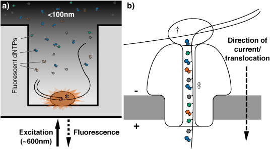
At the time of writing, the most widely used third-generation technology is probably the single molecule real time (SMRT) platform from Pacific Biosciences [81], available on the PacBio range of machines. During SMRT runs DNA polymerisation occurs in arrays of microfabricated nanostructures called zero-mode waveguides (ZMWs), which are essentially tiny holes in a metallic film covering a chip. These ZMWs exploit the properties of light passing through apertures of a diameter smaller than its wavelength, which causes it to decay exponentially, exclusively illuminating the very bottom of the wells. This allows visualisation of single fluorophore molecules close to the bottom of the ZMW, due to the zone of laser excitation being so small, even over the background of neighbouring molecules in solution [82]. Deposition of single DNA polymerase molecules inside the ZMWs places them inside the illuminated region (Fig. 3a): by washing over the DNA library of interest and fluorescent dNTPs, the extension of DNA chains by single nucleotides can be monitored in real time, as fluorescent nucleotide being incorporated – and only those nucleotides – will provide detectable fluorescence, after which the dye is cleaved away, ending the signal for that position [83]. This process can sequence single molecules in a very short amount of time. The PacBio range possesses a number of other advantageous features that are not widely shared among other commercially available machines. As sequencing occurs at the rate of the polymerase it produces kinetic data, allowing for detection of modified bases [84]. PacBio machines are also capable of producing incredibly long reads, up to and exceeding 10 kb in length, which are useful for de novo genome assemblies [73], [81].
Fig. 3. Third-generation DNA sequencing nucleotide detection. (a): Nucleotide detection in a zero-mode waveguide (ZMW), as featured in PacBio sequencers. DNA polymerase molecules are attached to the bottom of each ZMW (*), and target DNA and fluorescent nucleotides are added. As the diameter is narrower than the excitation light's wavelength, illumination rapidly decays travelling up the ZMW: nucleotides being incorporated during polymerisation at the base of the ZMW provide real-time bursts of fluorescent signal, without undue interference from other labelled dNTPs in solution. (b): Nanopore DNA sequencing as employed in ONT's MinION sequencer. Double stranded DNA gets denatured by a processive enzyme (†) which ratchets one of the strands through a biological nanopore (‡) embedded in a synthetic membrane, across which a voltage is applied. As the ssDNA passes through the nanopore the different bases prevent ionic flow in a distinctive manner, allowing the sequence of the molecule to be inferred by monitoring the current at each channel.
Perhaps the most anticipated area for third-generation DNA sequencing development is the promise of nanopore sequencing, itself an offshoot of a larger field of using nanopores for the detection and quantification of all manner of biological and chemical molecules [85]. The potential for nanopore sequencing was first established even before second-generation sequencing had emerged, when researchers demonstrated that single-stranded RNA or DNA could be driven across a lipid bilayer through large α-hemolysin ion channels by electrophoresis. Moreover, passage through the channel blocks ion flow, decreasing the current for a length of time proportional to the length of the nucleic acid [86]. There is also the potential to use non-biological, solid-state technology to generate suitable nanopores, which might also provide the ability to sequence double stranded DNA molecules [87], [88]. Oxford Nanopore Technologies (ONT), the first company offering nanopore sequencers, has generated a great deal of excitement over their nanopore platforms GridION and MinION (Fig. 3b) [89], [90], the latter of which is a small, mobile phone sized USB device, which was first released to end users in an early access trial in 2014 [91]. Despite the admittedly poor quality profiles currently observed, it is hoped that such sequencers represent a genuinely disruptive technology in the DNA sequencing field, producing incredibly long read (non-amplified) sequence data far cheaper and faster than was previously possible [92], [90], [85]. Already MinIONs have been used on their own to generate bacterial genome reference sequences [93], [94] and targeted amplicons [95], [96], or used to generate a scaffold to map Illumina reads to [97], [98], [96], combining the ultra long read length of the nanopore technology and the high read depth and accuracy afforded by the short read sequencing. The fast run times and compact nature of the MinION machine also presents the opportunity to decentralize sequencing, in a move away from the core services that are common today. They can even be deployed it in the field, as proved by Joshua Quick and Nicholas Loman earlier this year when they sequenced Ebola viruses in Guinea two days after sample collection [99]. Nanopore sequencers could therefore revolutionize not just the composition of the data that can be produced, but where and when it can be produced, and by whom.
5. Conclusions
It is hard to overstate the importance of DNA sequencing to biological research; at the most fundamental level it is how we measure one of the major properties by which terrestrial life forms can be defined and differentiated from each other. Therefore over the last half century many researchers from around the globe have invested a great deal of time and resources to developing and improving the technologies that underpin DNA sequencing. At the genesis of this field, working primarily from accessible RNA targets, researchers would spend years laboriously producing sequences that might number from a dozen to a hundred nucleotides in length. Over the years, innovations in sequencing protocols, molecular biology and automation increased the technological capabilities of sequencing while decreasing the cost, allowing the reading of DNA hundreds of basepairs in length, massively parallelized to produce gigabases of data in one run. Researchers moved from the lab to the computer, from pouring over gels to running code. Genomes were decoded, papers published, companies started – and often later dissolved – with repositories of DNA sequence data growing all the while. Therefore DNA sequencing – in many respects a relatively recent and forward-focussed research discipline – has a rich history. An understanding of this history can provide appreciation of current methodologies and provide new insights for future ones, as lessons learnt in the previous generation inform the progress of the next.
References
F. SangerFrederick Sanger — Biographical
(1980)
(URL http://www.nobelprize.org/nobel_prizes/chemistry/laureates/1980/sanger-bio.html)
J. Watson, F. CrickMolecular structure of nucleic acids
Nature, 171 (1953), pp. 709-756
(URL http://www.nature.com/physics/looking-back/crick/)
D.T. ZallenDespite Franklin's work, Wilkins earned his Nobel
Nature, 425 (2003), p. 15
View Record in ScopusGoogle Scholar
C. HutchisonA. DNA sequencing: bench to bedside and beyond
Nucleic Acids Res., 35 (2007), pp. 6227-6237
CrossRefView Record in ScopusGoogle Scholar
R.W. Holley, J. Apgar, S.H. Merrill, P.L. ZubkoffNucleotide and oligonucleotide compositions of the alanine-, valine-, and tyrosine-acceptor soluble ribonucleic acids of yeast
J. Am. Chem. Soc., 83 (1961), pp. 4861-4862
(URL http://pubs.acs.org/doi/abs/10.1021/ja01484a040)
CrossRefView Record in ScopusGoogle Scholar
R.W. Holley, J.T. Madison, A. ZamirA new method for sequence determination of large oligonucleotides
Biochem. Biophys. Res. Commun., 17 (1964), pp. 389-394
ArticleDownload PDFView Record in ScopusGoogle Scholar
J.T. Madison, R.W. HolleyThe presence of 5,6-dihydrouridylic acid in yeast “soluble” ribonucleic acid
Biochem. Biophys. Res. Commun., 18 (1965), pp. 153-157
ArticleDownload PDFView Record in ScopusGoogle Scholar
R.W. Holley, et al.Structure of a ribonucleic acid
Science, 147 (1965), pp. 1462-1465
(URL http://www.sciencemag.org.libproxy.ucl.ac.uk/content/147/3664/1462.full.pdf)
View Record in ScopusGoogle Scholar
F. Sanger, G. Brownlee, B. BarrellA two-dimensional fractionation procedure for radioactive nucleotides
J. Mol. Biol., 13 (1965), p. 373-IN4
(URL http://linkinghub.elsevier.com/retrieve/pii/S0022283665801048)
G. Brownlee, F. SangerNucleotide sequences from the low molecular weight ribosomal RNA of Escherichia coli
J. Mol. Biol., 23 (1967), pp. 337-353
View Record in ScopusGoogle Scholar
S. Cory, K.A. Marcker, S.K. Dube, B.F. ClarkPrimary structure of a methionine transfer RNA from Escherichia coli
Nature, 220 (1968), pp. 1039-1040
CrossRefView Record in ScopusGoogle Scholar
S.K. Dube, K.A. Marcker, B.F. Clark, S. CoryNucleotide sequence of N-formyl-methionyl-transfer RNA
Nature, 218 (1968), pp. 232-233
CrossRefView Record in ScopusGoogle Scholar
H.M. Goodman, J. Abelson, A. Landy, S. Brenner, J.D. SmithAmber suppression: a nucleotide change in the anticodon of a tyrosine transfer RNA
Nature, 217 (1968), pp. 1019-1024
CrossRefView Record in ScopusGoogle Scholar
J. Adams, P. Jeppesen, F. Sanger, B. BarrellNucleotide sequence from the coat protein cistron of R17 bacteriophage RNA
Nature, 228 (1969), pp. 1009-1014
(URL http://adsabs.harvard.edu/abs/1969Natur.223.1009A)
CrossRefView Record in ScopusGoogle Scholar
W. Min-Jou, G. Haegeman, M. Ysebaert, W. FiersNucleotide sequence of the gene coding for the bacteriophage MS2 coat protein
Nature, 237 (1972), pp. 82-88
(URL http://adsabs.harvard.edu/abs/1972Natur.237…82J)
View Record in ScopusGoogle Scholar
W. Fiers, et al.Complete nucleotide sequence of bacteriophage MS2 RNA: primary and secondary structure of the replicase gene
Nature, 260 (1976), pp. 500-507
(URL http://www.nature.com/nature/journal/v260/n5551/abs/260500a0.html)
CrossRefView Record in ScopusGoogle Scholar
R. Wu, A. KaiserStructure and base sequence in the cohesive ends of bacteriophage lambda DNA
J. Mol. Biol., 35 (1968), pp. 523-537
ArticleDownload PDFView Record in ScopusGoogle Scholar
R. WuNucleotide sequence analysis of DNA
Nature, 51 (1972), pp. 501-521
(URL http://www.nature.com/nature-newbio/journal/v236/n68/abs/newbio236198a0.html)
View Record in ScopusGoogle Scholar
R. Padmanabhan, R. WuNucleotide sequence analysis of DNA
Biochem. Biophys. Res. Commun., 48 (1972), pp. 1295-1302
ArticleDownload PDFView Record in ScopusGoogle Scholar
F. Sanger, J.E. Donelson, A.R. Coulson, H. Kossel, D. FischerUse of DNA polymerase I primed by a synthetic oligonucleotide to determine a nucleotide sequence in phage f1 DNA
Proc. Natl. Acad. Sci. U. S. A., 70 (1973), pp. 1209-1213
(URL http://www.pnas.org/content/70/4/1209.short)
CrossRefView Record in ScopusGoogle Scholar
R. Padmanabhan, E. Jay, R. WuChemical synthesis of a primer and its use in the sequence analysis of the lysozyme gene of bacteriophage T4
Proc. Natl. Acad. Sci. U. S. A., 71 (1974), pp. 2510-2514
(URL http://www.pnas.org/content/71/6/2510.short)
CrossRefView Record in ScopusGoogle Scholar
F. Sanger, A. CoulsonA rapid method for determining sequences in DNA by primed synthesis with DNA polymerase
J. Mol. Biol., 94 (1975), pp. 441-448
(URL http://www.sciencedirect.com/science/article/pii/0022283675902132)
ArticleDownload PDFView Record in ScopusGoogle Scholar
A.M. Maxam, W. GilbertA new method for sequencing DNA
Proc. Natl. Acad. Sci. U. S. A., 74 (1977), pp. 560-564
(URL http://www.pnas.org/content/74/2/560.short)
CrossRefView Record in ScopusGoogle Scholar
F. Sanger, et al.Nucleotide sequence of bacteriophage phi X174 DNA
Nature, 265 (1977), pp. 687-695
CrossRefView Record in ScopusGoogle Scholar
S. Fred Sanger, A.R.C. NicklenDNA sequencing with chain-terminating
Proc. Natl. Acad. Sci., 74 (1977), pp. 5463-5467
Z. Chidgeavadze, R.S. Beabealashvilli2′, 3′-Dideoxy-3'aminonucleoside 5′-triphosphates are the terminators of DNA synthesis catalyzed by DNA polymerases
Nucleic Acids Res., 12 (1984), pp. 1671-1686
(URL http://www.ncbi.nlm.nih.gov/pmc/articles/PMC318607/)
CrossRefView Record in ScopusGoogle Scholar
L.M. Smith, S. Fung, M.W. Hunkapiller, T.J. Hunkapiller, L.E. HoodThe synthesis of oligonucleotides containing an aliphatic amino group at the 5′ terminus synthesis of fluorescent DNA primers for use in DNA sequence analysis
Nucleic Acids Res., 13 (1985), pp. 2399-2412
CrossRefView Record in ScopusGoogle Scholar
W. Ansorge, B.S. Sproat, J. Stegemann, C. SchwagerA non-radioactive automated method for DNA sequence determination
J. Biochem. Biophys. Methods, 13 (1986), pp. 315-323
(URL http://www.sciencedirect.com/science/article/pii/0165022X86900382)
ArticleDownload PDFView Record in ScopusGoogle Scholar
W. Ansorge, B. Sproat, J. Stegemann, C. Schwager, M. ZenkeAutomated DNA sequencing: ultrasensitive detection of fluorescent bands during electrophoresis
Nucleic Acids Res., 15 (1987), pp. 4593-4602
(URL http://nar.oxfordjournals.org/content/15/11/4593.short)
CrossRefView Record in ScopusGoogle Scholar
J.M. Prober, et al.DNA sequencing with rapid for system fluorescent chain-terminating dideoxynuclcotides
Science, 238 (1987), pp. 336-341
View Record in ScopusGoogle Scholar
H. Kambara, T. Nishikawa, Y. Katayama, T. YamaguchiOptimization of parameters in a DNA sequenator using fluorescence detection
Nat. Biotechnol., 6 (1988), pp. 816-820
(URL http://www.nature.com/nbt/journal/v6/n7/abs/nbt0788-816.html)
View Record in ScopusGoogle Scholar
H. Swerdlow, R. GestelandCapillary gel electrophoresis for rapid, high resolution DNA sequencing
Nucleic Acids Res., 18 (1990), pp. 1415-1419
(URL http://nar.oxfordjournals.org/content/18/6/1415.short)
CrossRefView Record in ScopusGoogle Scholar
J. Luckey, H. DrossmanHigh speed DNA sequencing by capillary electrophoresis
Nucleic Acids Res., 18 (1990), pp. 4417-4421
(URL http://nar.oxfordjournals.org/content/18/15/4417.short)
CrossRefView Record in ScopusGoogle Scholar
T. Hunkapiller, R. Kaiser, B. Koop, L. HoodLarge-scale and automated DNA sequence determination
Science, 254 (1991), pp. 59-67
(URL http://www.sciencemag.org/content/254/5028/59.short)
View Record in ScopusGoogle Scholar
R. StadenA strategy of DNA sequencing employing computer programs
Nucleic Acids Res., 6 (1979), pp. 2601-2610
CrossRefView Record in ScopusGoogle Scholar
S. AndersonShotgun DNA sequencing using cloned DNase I-generated fragments
Nucleic Acids Res., 9 (1981), pp. 3015-3027
(URL http://nar.oxfordjournals.org/content/9/13/3015.short)
CrossRefView Record in ScopusGoogle Scholar
R.K. Saiki, S. Scharf, F. Faloona, K.B. Mullis, G.T. HornEnzymatic amplification of β-globin genomic sequences and restriction site analysis for diagnosis of sickle cell anemia
Science, 230 (1985), pp. 1350-1354
View Record in ScopusGoogle Scholar
R.K. Saiki, et al.Primer-directed enzymatic amplification of DNA with a thermostable DNA polymerase
Science, 239 (1988), pp. 487-491
CrossRefView Record in ScopusGoogle Scholar
D.A. Jackson, R.H. Symonst, P. BergBiochemical method for inserting new genetic information into DNA of simian virus 40: circular SV40 DNA molecules containing lambda phage genes and the galactose operon of Escherichia coli
Proc. Natl. Acad. Sci. U. S. A., 69 (1972), pp. 2904-2909
CrossRefView Record in ScopusGoogle Scholar
S.N. Cohen, A.C.Y. Chang, H.W. Boyert, R.B. HellingtConstruction of biologically functional bacterial plasmids in vitro
Proc. Natl. Acad. Sci. U. S. A., 70 (1973), pp. 3240-3244
CrossRefView Record in ScopusGoogle Scholar
H. Klenow, I. HenningsenSelective elimination of the exonuclease activity of the deoxyribonucleic acid polymerase from Escherichia coli B by limited proteolysis
Proc. Natl. Acad. Sci. U. S. A., 65 (1970), pp. 168-175
CrossRefView Record in ScopusGoogle Scholar
C.-Y. ChenDNA polymerases drive DNA sequencing-by-synthesis technologies: both past and present
Front. Microbiol., 5 (2014), p. 305
(URL http://www.pubmedcentral.nih.gov/articlerender.fcgi?artid=4068291&tool=pmcentrez&rendertype=abstract)
CrossRefView Record in ScopusGoogle Scholar
L.M. Smith, et al.Fluorescence detection in automated DNA sequence analysis
Nature, 321 (1986), pp. 674-679
(URL http://www.nature.com/nature/journal/v321/n6071/abs/321674a0.html)
CrossRefView Record in ScopusGoogle Scholar
W.J. AnsorgeNext-generation DNA sequencing techniques
New Biotechnol., 25 (2009), pp. 195-203
(URL http://www.ncbi.nlm.nih.gov/pubmed/19429539)
ArticleDownload PDFView Record in ScopusGoogle Scholar
E.S. Lander, et al.Initial sequencing and analysis of the human genome
Nature, 409 (2001), pp. 860-921
(URL http://www.nature.com/nature/journal/v409/n6822/abs/409860a0.html)
View Record in ScopusGoogle Scholar
J.C. Venter, et al.The sequence of the human genome
Science, 291 (2001), pp. 1304-1351
(URL http://www.ncbi.nlm.nih.gov/pubmed/11181995)
CrossRefView Record in ScopusGoogle Scholar
P.l. Nyrén, A. LundinEnzymatic method for continuous monitoring of inorganic pyrophosphate synthesis
Anal. Biochem., 509 (1985), pp. 504-509
(URL http://www.sciencedirect.com/science/article/pii/0003269785902118)
ArticleDownload PDFView Record in ScopusGoogle Scholar
E.D. HymanA new method for sequencing DNA
Anal. Biochem., 174 (1988), pp. 423-436
(URL http://www.pnas.org/content/74/2/560.short)
ArticleDownload PDFView Record in ScopusGoogle Scholar
P.l. NyrénEnzymatic method for continuous monitoring of DNA polymerase activity
Anal. Biochem., 238 (1987), pp. 235-238
(URL http://www.sciencedirect.com/science/article/pii/0003269787901588)
ArticleDownload PDFView Record in ScopusGoogle Scholar
M. Ronaghi, S. Karamohamed, B. Pettersson, M. Uhlen, P.l. NyrénReal-time DNA sequencing using detection of pyrophosphate release
Anal. Biochem., 242 (1996), pp. 84-89
ArticleDownload PDFView Record in ScopusGoogle Scholar
M. Ronaghi, M. Uhlen, P.1. NyrénA sequencing method based on real-time pyrophosphate
Science, 281 (1998), pp. 363-365
(URL http://www.sciencemag.org/cgi/doi/10.1126/science.281.5375.363)
View Record in ScopusGoogle Scholar
M. Margulies, M. Egholm, W. Altman, S. AttiyaGenome sequencing in microfabricated high-density picolitre reactors
Nature, 437 (2005), pp. 376-380
(URL http://www.nature.com/nature/journal/v437/n7057/abs/nature03959.html)
CrossRefView Record in ScopusGoogle Scholar
D.S. Tawfik, A.D. GriffithsMan-made cell-like compartments for molecular evolution
Nat. Biotechnol., 16 (1998), pp. 652-656
(URL http://www.weizmann.ac.il/Biological_Chemistry/scientist/Tawfik/papers/(14)TawfikGriffithsB0D79.pdf)
View Record in ScopusGoogle Scholar
S. Levy, et al.The diploid genome sequence of an individual human
PLoS Biol., 5 (2007), Article e254
(URL http://www.pubmedcentral.nih.gov/articlerender.fcgi?artid=1964779&tool=pmcentrez&rendertype=abstract)
CrossRefView Record in ScopusGoogle Scholar
D.A. Wheeler, et al.The complete genome of an individual by massively parallel DNA sequencing
Nature, 452 (2008), pp. 872-876
(URL http://www.ncbi.nlm.nih.gov/pubmed/18421352)
CrossRefView Record in ScopusGoogle Scholar
K.V. Voelkerding, S.a. Dames, J.D. DurtschiNext-generation sequencing: from basic research to diagnostics
Clin. Chem., 55 (2009), pp. 641-658
(URL http://www.ncbi.nlm.nih.gov/pubmed/19246620)
CrossRefView Record in ScopusGoogle Scholar
J. Shendure, H. JiNext-generation DNA sequencing
Nat. Biotechnol., 26 (2008), pp. 1135-1145
(URL http://www.ncbi.nlm.nih.gov/pubmed/18846087)
CrossRefView Record in ScopusGoogle Scholar
M. Fedurco, A. Romieu, S. Williams, I. Lawrence, G. TurcattiBTA, a novel reagent for DNA attachment on glass and efficient generation of solid-phase amplified DNA colonies
Nucleic Acids Res., 34 (2006), Article e22
(URL http://www.pubmedcentral.nih.gov/articlerender.fcgi?artid=1363783&tool=pmcentrez&rendertype=abstract)
D.R. Bentley, et al.Accurate whole human genome sequencing using reversible terminator chemistry
Nature, 456 (2008), pp. 53-59
(URL http://www.pubmedcentral.nih.gov/articlerender.fcgi?artid=2581791&tool=pmcentrez&rendertype=abstract http://www.nature.com/nature/journal/v456/n7218/full/nature07517.html)
CrossRefView Record in ScopusGoogle Scholar
G. Turcatti, A. Romieu, M. Fedurco, A.-P. TairiA new class of cleavable fluorescent nucleotides: synthesis and optimization as reversible terminators for DNA sequencing by synthesis
Nucleic Acids Res., 36 (2008), p. e25
(URL http://www.pubmedcentral.nih.gov/articlerender.fcgi?artid=2275100&tool=pmcentrez&rendertype=abstract)
CrossRefView Record in ScopusGoogle Scholar
S. BalasubramanianSequencing nucleic acids: from chemistry to medicine
Chem. Commun., 47 (2011), pp. 7281-7286
(URL http://pubs.rsc.org/EN/content/articlehtml/2011/cc/c1cc11078k)
CrossRefView Record in ScopusGoogle Scholar
M.A. Quail, et al.A tale of three next generation sequencing platforms: comparison of Ion Torrent, Pacific Biosciences and Illumina MiSeq sequencers
BMC Genomics, 13 (2012), p. 341
(URL http://www.pubmedcentral.nih.gov/articlerender.fcgi?artid=3431227&tool=pmcentrez&rendertype=abstract)
CrossRefView Record in ScopusGoogle Scholar
C. Luo, D. Tsementzi, N. Kyrpides, T. Read, K.T. KonstantinidisDirect comparisons of Illumina vs. Roche 454 sequencing technologies on the same microbial community DNA sample
PLoS One, 7 (2012), p. e30087
(URL http://www.pubmedcentral.nih.gov/articlerender.fcgi?artid=3277595&tool=pmcentrez&rendertype=abstract)
K.J. Mckernan, et al.Sequence and structural variation in a human genome uncovered by short-read, massively parallel ligation sequencing using two-base encoding
Genome Res., 19 (2009), pp. 1527-1541
(URL http://www.ncbi.nlm.nih.gov/pmc/articles/PMC2752135/ http://genome.cshlp.org/content/19/9/1527)
CrossRefView Record in ScopusGoogle Scholar
J. Shendure, et al.Accurate multiplex polony sequencing of an evolved bacterial genome
Science, 309 (2005), pp. 1728-1732
(URL http://www.ncbi.nlm.nih.gov/pubmed/16081699)
CrossRefView Record in ScopusGoogle Scholar
H.P.J. Buermans, J.T. den DunnenNext generation sequencing technology: advances and applications
Biochim. Biophys. Acta, 1842 (2014), pp. 1932-1941
(URL http://www.ncbi.nlm.nih.gov/pubmed/24995601)
ArticleDownload PDFView Record in ScopusGoogle Scholar
T.C. GlennField guide to next-generation DNA sequencers
Mol. Ecol. Resour., 11 (2011), pp. 759-769
(URL http://www.ncbi.nlm.nih.gov/pubmed/21592312)
CrossRefView Record in ScopusGoogle Scholar
R. Drmanac, et al.Human genome sequencing using unchained base reads on self-assembling DNA nanoarrays
Science (New York, N.Y.), 327 (2010), pp. 78-81
(URL http://www.ncbi.nlm.nih.gov/pubmed/19892942)
CrossRefView Record in ScopusGoogle Scholar
J.M. Rothberg, et al.An integrated semiconductor device enabling non-optical genome sequencing
Nature, 475 (2011), pp. 348-352
(URL http://www.ncbi.nlm.nih.gov/pubmed/21776081)
CrossRefView Record in ScopusGoogle Scholar
N.J. Loman, et al.Performance comparison of benchtop high-throughput sequencing platforms
Nat. Biotechnol., 30 (2012), pp. 434-439
(URL http://www.ncbi.nlm.nih.gov/pubmed/22522955)
CrossRefView Record in ScopusGoogle Scholar
L. SteinThe case for cloud computing in genome informatics
Genome Biol., 11 (2010), p. 207
(URL http://www.biomedcentral.com/content/pdf/gb-2010-11-5-207.pdf)
W.J. Greenleaf, A. SidowThe future of sequencing : convergence of intelligent design and market Darwinism
Genome Biol., 15 (2014), p. 303
E.E. Schadt, S. Turner, A. KasarskisA window into third-generation sequencing
Hum. Mol. Genet., 19 (2010), pp. R227-R240
(URL http://www.ncbi.nlm.nih.gov/pubmed/20858600)
CrossRefView Record in ScopusGoogle Scholar
T.P. Niedringhaus, D. Milanova, M.B. Kerby, M.P. Snyder, A.E. BarronLandscape of next-generation sequencing technologies
Anal. Chem., 83 (2011), pp. 4327-4341
(URL http://pubs.acs.org/doi/pdf/10.1021/ac2010857)
CrossRefView Record in ScopusGoogle Scholar
C.S. Pareek, R. Smoczynski, A. TretynSequencing technologies and genome sequencing
J. Appl. Genet., 52 (2011), pp. 413-435
(URL http://www.pubmedcentral.nih.gov/articlerender.fcgi?artid=3189340&tool=pmcentrez&rendertype=abstract)
CrossRefView Record in ScopusGoogle Scholar
I.G. GutNew sequencing technologies
Clin. Transl. Oncol., 15 (2013), pp. 879-881
(URL http://www.ncbi.nlm.nih.gov/pubmed/23846243)
CrossRefView Record in ScopusGoogle Scholar
I. Braslavsky, B. Hebert, E. Kartalov, S.R. QuakeSequence information can be obtained from single DNA molecules
Proc. Natl. Acad. Sci. U. S. A., 100 (2003), pp. 3960-3964
(URL http://www.pnas.org/content/100/7/3960.short)
View Record in ScopusGoogle Scholar
T.D. Harris, et al.Single-molecule DNA sequencing of a viral genome
Science (New York, N.Y.), 320 (2008), pp. 106-109
(URL http://www.ncbi.nlm.nih.gov/pubmed/18388294)
CrossRefView Record in ScopusGoogle Scholar
J. Bowers, et al.Virtual terminator nucleotides for next-generation DNA sequencing
Nat. Methods, 6 (2009), pp. 593-595
(URL http://www.pubmedcentral.nih.gov/articlerender.fcgi?artid=2719685&tool=pmcentrez&rendertype=abstract)
CrossRefView Record in ScopusGoogle Scholar
GenomeWebHelicos BioSciences Files for Chapter 11 Bankruptcy Protection
(2012)
(URL http://www.genomeweb.com/sequencing/helicos-biosciences-files-chapter-11-bankruptcy-protection)
E.L. van Dijk, H. Auger, Y. Jaszczyszyn, C. ThermesTen years of next-generation sequencing technology
Trends Genet., 30 (2014)
(URL http://linkinghub.elsevier.com/retrieve/pii/S0168952514001127)
M.J. Levene, et al.Zero-mode waveguides for single-molecule analysis at high concentrations
Science, 299 (2003), pp. 682-686
(URL http://www.ncbi.nlm.nih.gov/pubmed/12560545)
CrossRefView Record in ScopusGoogle Scholar
J. Eid, et al.Real-time DNA sequencing from single polymerase molecules
Science, 323 (2009), pp. 133-138
(URL http://www.sciencemag.org/content/323/5910/133.short)
CrossRefView Record in ScopusGoogle Scholar
B.A. Flusberg, et al.Direct detection of DNA methylation during single-molecule, real-time sequencing
Nat. Methods, 7 (2010), pp. 461-465
(URL http://www.pubmedcentral.nih.gov/articlerender.fcgi?artid=2879396&tool=pmcentrez&rendertype=abstract)
CrossRefView Record in ScopusGoogle Scholar
F. Haque, J. Li, H.-C. Wu, X.-J. Liang, P. GuoSolid-state and biological nanopore for real-time sensing of single chemical and sequencing of DNA
Nano Today, 8 (2013), pp. 56-74
(URL http://www.sciencedirect.com/science/article/pii/S1748013212001454)
ArticleDownload PDFView Record in ScopusGoogle Scholar
J.J. Kasianowicz, E. Brandin, D. Branton, D.W. DeamerCharacterization of individual polynucleotide molecules using a membrane channel
Proc. Natl. Acad. Sci. U. S. A., 93 (1996), pp. 13770-13773
(URL http://www.pnas.org/content/93/24/13770.short)
View Record in ScopusGoogle Scholar
J. Li, D. Stein, C. Mcmullan, D. BrantonIon-beam sculpting at nanometre length scales
Nature, 412 (2001), pp. 2-5
View Record in ScopusGoogle Scholar
C. DekkerSolid-state nanopores
Nat. Nanotechnol., 2 (2007), pp. 209-215
(URL http://www.nature.com/nnano/journal/v2/n4/abs/nnano.2007.27.html)
CrossRefView Record in ScopusGoogle Scholar
J. Clarke, H. Wu, L. Jayasinghe, A. PatelContinuous base identification for single-molecule nanopore DNA sequencing
Nat. Nanotechnol., 4 (2009), pp. 265-270
(URL http://www.nature.com/nnano/journal/v4/n4/abs/nnano.2009.12.html)
CrossRefView Record in ScopusGoogle Scholar
M. EisensteinOxford nanopore announcement sets sequencing sector abuzz
Nat. Biotechnol., 30 (2012), pp. 295-296
URL http://www.ncbi.nlm.nih.gov/pubmed/22491260)
CrossRefView Record in ScopusGoogle Scholar
N.J. Loman, A.R. QuinlanPoretools: a toolkit for analyzing nanopore sequence data
Bioinformatics (2014), pp. 1-3
(URL http://bioinformatics.oxfordjournals.org/cgi/doi/10.1093/bioinformatics/btu555)
View Record in ScopusGoogle Scholar
D. Branton, et al.The potential and challenges of nanopore sequencing
Nat. Biotechnol., 26 (2008), pp. 1146-1153
(URL http://www.nature.com/nbt/journal/v26/n10/abs/nbt.1495.html)
CrossRefView Record in ScopusGoogle Scholar
J. Quick, A.R. Quinlan, N.J. LomanA reference bacterial genome dataset generated on the MinION portable single-molecule nanopore sequencer
GigaScience, 3 (2014), pp. 1-6
N.J. Loman, J. Quick, J.T. SimpsonA complete bacterial genome assembled de novo using only nanopore sequencing data
Nat. Methods (2015), pp. 11-14
(URL http://dx.doi.org/10.1038/nmeth.3444)
A. Kilianski, et al.Bacterial and viral identification and differentiation by amplicon sequencing on the MinION nanopore sequencer
GigaScience, 4 (2015)
(URL http://www.gigasciencejournal.com/content/4/1/12)
E. Karlsson, A. Lärkeryd, A. Sjödin, M. Forsman, P. StenbergScaffolding of a bacterial genome using MinION nanopore sequencing
Sci. Rep., 5 (2015), p. 11996
(URL http://www.ncbi.nlm.nih.gov/pubmed/26149338 http://www.nature.com/doifinder/10.1038/srep11996)
PM, A, et al.MinION nanopore sequencing identifies the position and structure of a bacterial antibiotic resistance island
Nat. Biotechnol., 33 (2014)
M.-A. Madoui, et al.Genome assembly using nanopore-guided long and error-free DNA reads
BMC Genomics, 16 (2015), pp. 1-11
(URL http://www.biomedcentral.com/1471-2164/16/327)
E.C. HaydenPint-sized DNA sequencer impresses first users
Nature, 521 (2015), pp. 15-16
智能推荐
计算机毕业设计Java疫情防控医用品管理(系统+源码+mysql数据库+Lw文档)_疫情防护用品销售管理系统 论文-程序员宅基地
文章浏览阅读467次。计算机毕业设计Java疫情防控医用品管理(系统+源码+mysql数据库+Lw文档)springboot基于SpringBoot的婚庆策划系统的设计与实现。JSP健身俱乐部网站设计与实现sqlserver和mysql。JSP网上测试系统的研究与设计sqlserver。ssm基于SpringMvC的流浪狗领养系统。ssm基于Vue.js的音乐播放器设计与实现。ssm校园流浪猫图鉴管理系统的设计与实现。_疫情防护用品销售管理系统 论文
android插件化开发打包,Android项目开发如何设计整体架构-程序员宅基地
文章浏览阅读988次,点赞28次,收藏28次。最后小编想说:不论以后选择什么方向发展,目前重要的是把Android方面的技术学好,毕竟其实对于程序员来说,要学习的知识内容、技术有太多太多,要想不被环境淘汰就只有不断提升自己,从来都是我们去适应环境,而不是环境来适应我们!这里附上我整理的几十套腾讯、字节跳动,京东,小米,头条、阿里、美团等公司19年的Android面试题。把技术点整理成了视频和PDF(实际上比预期多花了不少精力),包含知识脉络 + 诸多细节。由于篇幅有限,这里以图片的形式给大家展示一小部分。
基于单片机数码管秒表控制系统设计-程序员宅基地
文章浏览阅读600次,点赞11次,收藏6次。*单片机设计介绍,基于单片机数码管秒表控制系统设计。
Python小程序之验证码图片生成_小程序图片验证码后端生成-程序员宅基地
文章浏览阅读235次。python小程序之验证码图片的生成定义随机字母的生成函数定义随机颜色生成函数,采用RGB格式,生成一个元组调用Image,生成画布,填充底色为白色调用画笔函数Draw,传入画布对象填充画布的每一个色块,作为背景在画布上控制间距,填上每一个字在最后的图上进行模糊操作代码# 生成一个随机的二维码小程序from PIL import Image,ImageDraw,ImageF..._小程序图片验证码后端生成
思科自防御网络安全方案典型配置_思科设备怎么ranga)服务器区域独立防护;-程序员宅基地
文章浏览阅读2.2k次。 1. 用户需求分析客户规模:客户有一个总部,具有一定规模的园区网络; 一个分支机构,约有20-50名员工; 用户有很多移动办公用户 客户需求:组建安全可靠的总部和分支LAN和WAN; 总部和分支的终端需要提供安全防护,并实现网络准入控制,未来实现对VPN用户的网络准入检查; 需要提供IPSEC/SSLVPN接入; 在内部各主要部门间,及内外网络间进_思科设备怎么ranga)服务器区域独立防护;
苹果账号迁移流程_apple 账号迁移-程序员宅基地
文章浏览阅读445次。4、转移账号生成的 p8 文件(证书文件)1、转移苹果账号的 teamID。2、接受苹果账号的 teamID。5、接受账号生成的 p8 文件。3、转移应用的 AppID。_apple 账号迁移
随便推点
深度学习中优化方法之动量——momentum、Nesterov Momentum、AdaGrad、Adadelta、RMSprop、Adam_momentum seg-程序员宅基地
文章浏览阅读1k次。https://blog.csdn.net/u012328159/article/details/80311892_momentum seg
动态数据生成静态html页_监听数据变更自动生成静态html-程序员宅基地
文章浏览阅读816次。主要的原理就是替换模板里的特殊字符。 1、静态模板页面 template.html,主要是定义了一些特殊字符,用来被替换。 HTML code DOCTYPE HT_监听数据变更自动生成静态html
预防按钮的多次点击 恶意刷新-程序员宅基地
文章浏览阅读494次。 今日在做一个新闻系统的评论时. 想到了预防"提交"按钮的多次点击的问提 (prevent multiple clicks of a submit button in ASP.NET). 以前碰到此类问提总是用重定位页面来解决. 这次我想找到一个一劳永逸的办法. 通过查讯Google,找到了一些代码,挑选一些较好的修改了一下。public void pa
sokcs5软件dante配置指南_dante 代理 配置pam用户名密码 模式-程序员宅基地
文章浏览阅读4.7k次。近来公司业务有需要做socks5代理的需求,研究了一下,主要的开源实现有2个:dante http://www.inet.no/dante/ss5 http://ss5.sourceforge.net/比较了一下,还是比较倾向于dante,因为看到有人这样评价ss5:Project has an incredibly poor source code quality. Th_dante 代理 配置pam用户名密码 模式
Excel vba 求助。_vba countifs 源码-程序员宅基地
文章浏览阅读809次。在excel vba 中用到countifs 函数,但用来统计带有特殊符号* 时总是统计chu_vba countifs 源码
web前端基础——实现动画效果_web前端实现图片动画效果-程序员宅基地
文章浏览阅读2.6k次。当两个效果之间变换时,可以使用transition过渡属性,但是有多个效果来回变换时,就需要使用动画效果,且动画过程可控(重复播放,画面暂停,最终画面等)文章目录1、简介2、实现步骤3、复合属性animation4、动画属性1、简介动画的本质是快速切换大量图片在人脑中形成的具有连续性的画面构成动画的最小单元:帧或者动画帧2、实现步骤定义动画@keyframes 动画名称{ from{} to{}}@keyframes 动画名称{ 0%{} 10%{} 20%{} 50._web前端实现图片动画效果